Almost universal SERS sensor could change how we sniff out small things

sers

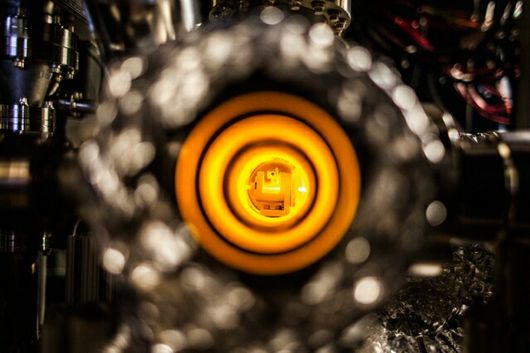
A new almost–universal SERS substrate could be the key to cheaper and easier sensors for drugs, explosives, or anything else (Credit: University of Buffalo)

Identifying fraudulent paintings based on electrochemical data, highlighting cancerous cells in a sea of healthy ones, and identifying different strains of bacteria in samples of food are all examples of surface-enhanced Raman spectroscopy (SERS), a sensor system that has only become more in-demand as our desire for precise, instantaneous information has increased. However, the technology has largely failed commercialization because the chips used are difficult and expensive to create, have limited uses for a particular known substance, and are consumed upon use. Researchers led by a team from the University of Buffalo (UB) aim to change nanoscale sensors with an almost-universal substrate that’s also low-cost, opening up more opportunities for powerful analysis of our environment.

Though SERS is complicated to understand on the surface, it forms a critical part of testing for explosives, identifying toxins in food, and other applications in public health and safety, medicine, and research.

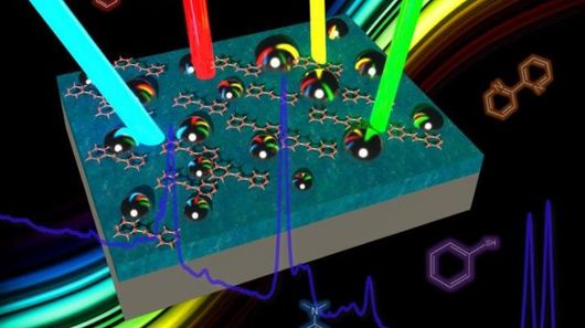
The technique relies on the unique electromagnetic properties of chemical compounds when stimulated with varying wavelengths of laser light and interacting with a surface designed to enhance the response (the “surface” in the name SERS). Each unique compound has a distinct spectral fingerprint and thus an industry researcher can discern between compounds that are invisible to the human eye without having to rely on doping the sample with labeling chemicals or having to possess a large sample.

Yet, currently most surfaces or substrates available on a commercial chip are optimized for only one wavelength of light, meaning scientists working with multiple compounds may need several chips to identify all their samples. This also ignores the ability to identify anonymous samples, which by their nature would require testing on multiple substrates.

The research from the team from UB and Fudan University in China introduces a substrate with a broadband nanostructure that “traps” the wide range of light most often used in SERS analysis, between 450 and 1100 nm.

The surface is composed of a film of thin silver or aluminum acting as a mirror, and a dielectric silica or alumina layer which separates the “mirror” from a layer of randomly applied silver nanoparticles. This construction also avoids expensive lithographic construction techniques.

Researcher Nan Zhang summed up the importance of the design by comparing it to a skeleton key.

“Instead of needing all these different substrates to measure Raman signals excited by different wavelengths, you’ll eventually need just one,” says Zhang. “Just like a skeleton key that opens many doors.”

Perhaps soon these “keys” will be available for airport screening, counterfeit protection, chemical weapon detection and a host of many more purposes requiring flexible, cheap sensors.

“The applications of such a device are far-reaching,” said Kai Liu, a PhD candidate in electrical engineering at UB. “The ability to detect even smaller amounts of chemical and biological molecules could be helpful with biosensors that are used to detect cancer, Malaria, HIV and other illnesses.”

References:http://www.gizmag.com/

MIT physicists build world’s first fermion microscope

fermion-microscope

Scientists at Massachusetts Institute of Technology have created a microscope that they claim is able to image the fundamental particles that make up all matter in the universe (Credit: Jose-Luis Olivares/MIT)

Researchers working at the Massachusetts Institute of Technology (MIT) claim to have created a method to better observe fermions – the sub-atomic building blocks of matter – by constructing a microscope capable of viewing them in groups of a thousand at a time. A laser technique is used to herd the fermions into a viewing area and then freeze them in place so all of the captured particles can be imaged simultaneously.

In the entire known universe, there are only two types of particles: fermions and bosons. In simple terms, fermions are all the particles that make up matter (for example, electrons), and bosons are all the particles that carry force (for example, photons).

Fermions include electrons, neutrons, quarks, protons, and atoms consisting of an odd number of any or all of these elementary particles. However, due to the strange (and not completely understood) nature of these particles in regard to their quantum spin states, scientists often opt to employ gases of ultra-cold fermionic atoms as proxies for     other fermions

.fermion-microscope-3

Over the last two decades, physicists studying ultracold atomic gases of boson particles – such as photons – have been able to do so relatively easily because bosons can occupy the same quantum state in boundless numbers. Fermions, however, are much harder to manipulate for imaging, as they are unable to be held in the same quantum state in large numbers and are very much more difficult to reduce to the temperatures required to slow them down enough to view them.

Physicists at Harvard University successfully created a boson microscope that could resolve individual bosons in an optical lattice as far back as 2009. Similarly, in 2010, the Max Planck Institute of Quantum Optics also developed a second boson microscope. And, though these microscopes exposed the behavior of bosons, their counterparts – fermions – remained elusive without an equivalent fermion microscope. .

“We wanted to do what these groups had done for bosons, but for fermions,” said Zwierlein says. “And it turned out it was much harder for fermions, because the atoms we use are not so easily cooled. So we had to find a new way to cool them while looking at them.”

What is required to study fermions is a way to reduce their temperature, and therefore their movement, to a point low enough to image them. However, even techniques that resulted in the first ever laboratory realization of Bose-Einstein condensation in 1995 (which resulted in a Nobel Prize in 2001), or later work that saw lasers cool atoms to a few ten-thousandths of a degree above absolute zero are insufficient to achieve the cooling required to image fermion atoms.

To overcome this problem, the MIT researchers initially created an optical lattice using laser beams to form an arrangement of light “wells” which could magnetically trap and hold a single fermion in place (a technique similar to that used by the University of California to capture cesium atoms and image rotons). Applying a number of stages of laser temperature reduction, and more evaporative cooling of the gas (in this case, potassium gas), the atoms were cooled to just above absolute zero which was cold enough to hold individual fermions in place on the optical lattice.

As the fermions move to this lower energy state, they also release photons of light which can then be captured by the microscope and used to locate a fermion’s exact position within the lattice at an accuracy level greater than the wavelength of light.

“That means I know where they are, and I can maybe move them around with a little tweezer to any location, and arrange them in any pattern I’d like,” said Martin Zwierlein, a professor of physics at MIT and a member of the team working on the project.

Unfortunately, this stability was tenuous because – when light was shone upon the atoms to view them – individual photons were able to knock them out of place.

.fermion-microscope-4

The team resolved this by cleverly employing a two laser beam approach where beams of differing frequencies were used to alter the fermion atom’s energy state. By simultaneously firing the two beams at the atom so that one beam frequency was absorbed by the particle, it would emit a corresponding photon in response. This, in turn, forced the particle into a lower energy state, thus cooling it further by reducing its excitation levels.

The upshot of this research, according to the team, is that the high-resolution image capture of more than 1,000 fermionic atoms all together at the one time will help improve our fundamental understanding of these elusive particles. As electrons are also fermions, it is hoped that this information may eventually aid research into high-temperature superconductors, with their inherent advantages of lossless energy transport and the development of quantum computer systems.

“The Fermi gas microscope, together with the ability to position atoms at will, might be an important step toward the realization of a quantum computer based on fermions,” said Zwierlein. “One would thus harness the power of the very same intricate quantum rules that so far hamper our understanding of electronic systems.”

References:http://www.gizmag.com/

Smartphone and tablet could be used for cheap, portable medical biosensing

smartphone-tablet-biosensing

A diagram of the CNBP system (Credit: Centre for Nanoscale BioPhotonics)

As mobile technology progresses, we’re seeing more and more examples of low-cost diagnostic systems being created for use in developing nations and remote locations. One of the latest incorporates little more than a smartphone, tablet, polarizer and box to test body fluid samples for diseases such as arthritis, cystic fibrosis and acute pancreatitis.

Developed at Australia’s Centre for Nanoscale BioPhotonics (CNBP), the setup utilizes fluorescent microscopy, a process in which dyes added to a sample cause specific biomarkers to glow when exposed to bright light.

To use it, clinicians deposit a dyed fluid sample in a well plate (basically a transparent sample-holding tray), put that plate on the screen of a tablet that’s in the box, and place a piece of polarizing glass over the plate compartment that contains the fluid. They then put their smartphone on top of the box, so that its camera lines up with that compartment.

Once the tablet is powered up, the light from its screen causes the targeted biomarkers to fluoresce (assuming they’re present in the first place). The polarizer allows light given off by those biomarkers to stand out from the tablet’s light, while an app on the phone analyzes the color and intensity of the fluorescence to help make a diagnosis.

“This type of fluorescent testing can be carried out by a variety of devices but in most cases the readout requires professional research laboratory equipment, which costs many tens of thousands of dollars,” says Ewa Goldys, CNBP’s deputy director. “What we’ve done is develop a device with a minimal number of commonly available components … The results can be analyzed by simply taking an image and the readout is available immediately.”

The free smartphone app will be available as of June 15th, via the project website. A paper on the research was recently published in the journal Sensors.

References:http://www.gizmag.com/

Researchers may have discovered fountain of youth by reversing aging in human cells

reverse-aging

Researchers in Japan have found that human aging may be able to be delayed or even reversed, at least at the most basic level of human cell lines. In the process, the scientists from the University of Tsukuba also found that regulation of two genes is related to how we age.

The new findings challenge one of the current popular theories of aging, that lays the blame for humans’ inevitable downhill slide with mutations that accumulate in our mitochondrial DNA over time. Mitochondrion are sometimes likened to a cellular “furnace” that produces energy through cellular respiration. Damage to the mitochondrial DNA results in changes or mutations in the DNA sequence that build up and are associated with familiar signs of aging like hair loss, osteoporosis and, of course, reduced lifespan.

So goes the theory, at least. But the Tsukuba researchers suggest that something else may be going on within our cells. Their research indicates that the issue may not be that mitochondrial DNA become damaged, but rather that genes get turned “off” or “on” over time. Most intriguing, the team led by Professor Jun-Ichi Hayashi was able to flip the switches on a few genes back to their youthful position, effectively reversing the aging process.

The researchers came to this conclusion by comparing the function level of the mitochondria in fibroblast cell lines from children under 12 years of age to those of elderly people between 80 and 97. As expected, the older cells had reduced cellular respiration, but the older cells did not show more DNA damage than those from children. This discovery led the team to propose that the reduced cellular function is tied to epigenetic regulation, changes that alter the physical structure of DNA without affecting the DNA sequence itself, causing genes to be turned on or off. Unlike mutations that damage that sequence, as in the other, aforementioned theory of aging, epigenetic changes could possibly be reversed by genetically reprogramming cells to an embryonic stem cell-like state, effectively turning back the clock on aging.

For a broad comparison, imagine that a power surge hits your home’s electrical system. If not properly wired, irreversible damage or even fire may result. However, imagine another home in which the same surge trips a switch in this home’s circuit breaker box. Simply flipping that breaker back to the “on” position should make it operate as good as new. In essence, the Tsukuba team is proposing that our DNA may not become fried with age as previously thought, but rather simply requires someone to access its genetic breaker box to reverse aging.

To test the theory, the researchers found two genes associated with mitochondrial function and essentially experimented with turning them on or off. In doing so, they were able to create defects or restore cellular respiration. These two genes regulate glycine, an amino acid, production in mitochondria, and in one of the more promising findings, a 97-year-old cell line saw its cellular respiration restored after the addition of glycine for 10 days.

The researchers’ findings were published this month in the journal Scientific Reports.

Whether or not this process could be a potential fountain of youth for humans and not just human fibroblast cell lines still remains to be seen, with much more testing required. However, if the theory holds, glycine supplements could one day become a powerful tool for life extension.

Similar research from the Salk Institute has also recently looked at other ways to slow down or stop aging at a cellular level, while yet another team is looking into a new class of drugs called senolytics that could help slow aging.

References:http://www.gizmag.com/